Healthcare
Kyoto University Hospital - Performance of EIZO Monitors Determines Our Reading Quality
The Department of Diagnostic Imaging and Nuclear Medicine at Kyoto University Hospital performs about 300 diagnostic image interpretations per day. In January 2022, they introduced 33 EIZO RadiForce RX660 monitors and two RX1270 monitors into their workflow for these interpretations. We interviewed Dr. Koji Fujimoto and Dr. Masako Kataoka from this department to hear their thoughts on using these monitors.
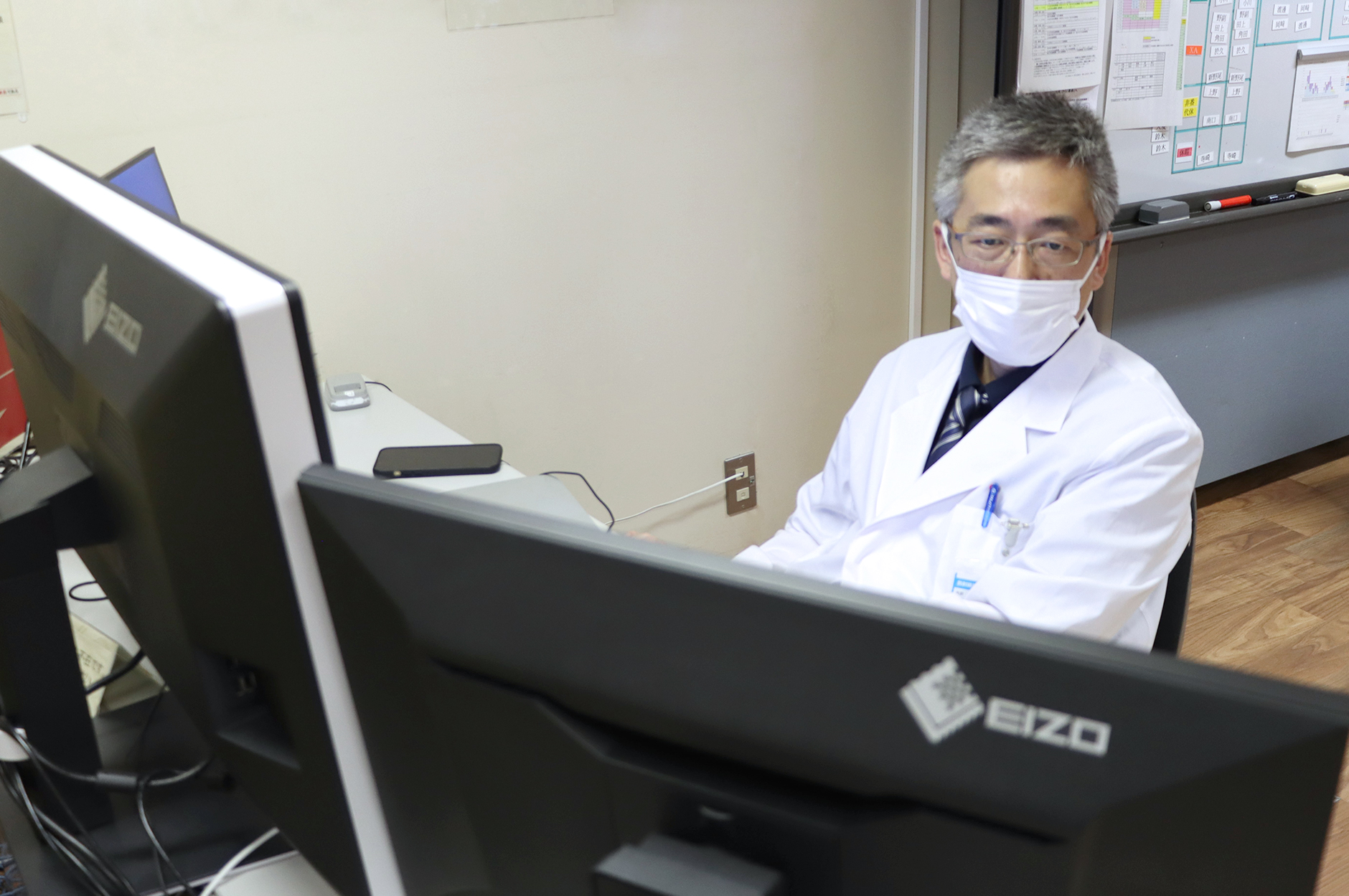
Dr. Koji Fujimoto |
Dr. Masako Kataoka |
What is the main function of the Department of Diagnostic Imaging and Nuclear Medicine?
The Importance of Consultations
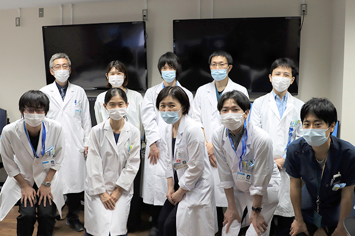
The hospital handles a significant volume of CT, MRI, and radioisotope (RI) scans. The Department of Diagnostic Imaging and Nuclear Medicine is responsible for performing an average of approximately 300 interpretations per day. It is common for more than ten staff members – or more than 20 people when graduate students, residents, and interns are included – to simultaneously review the images.
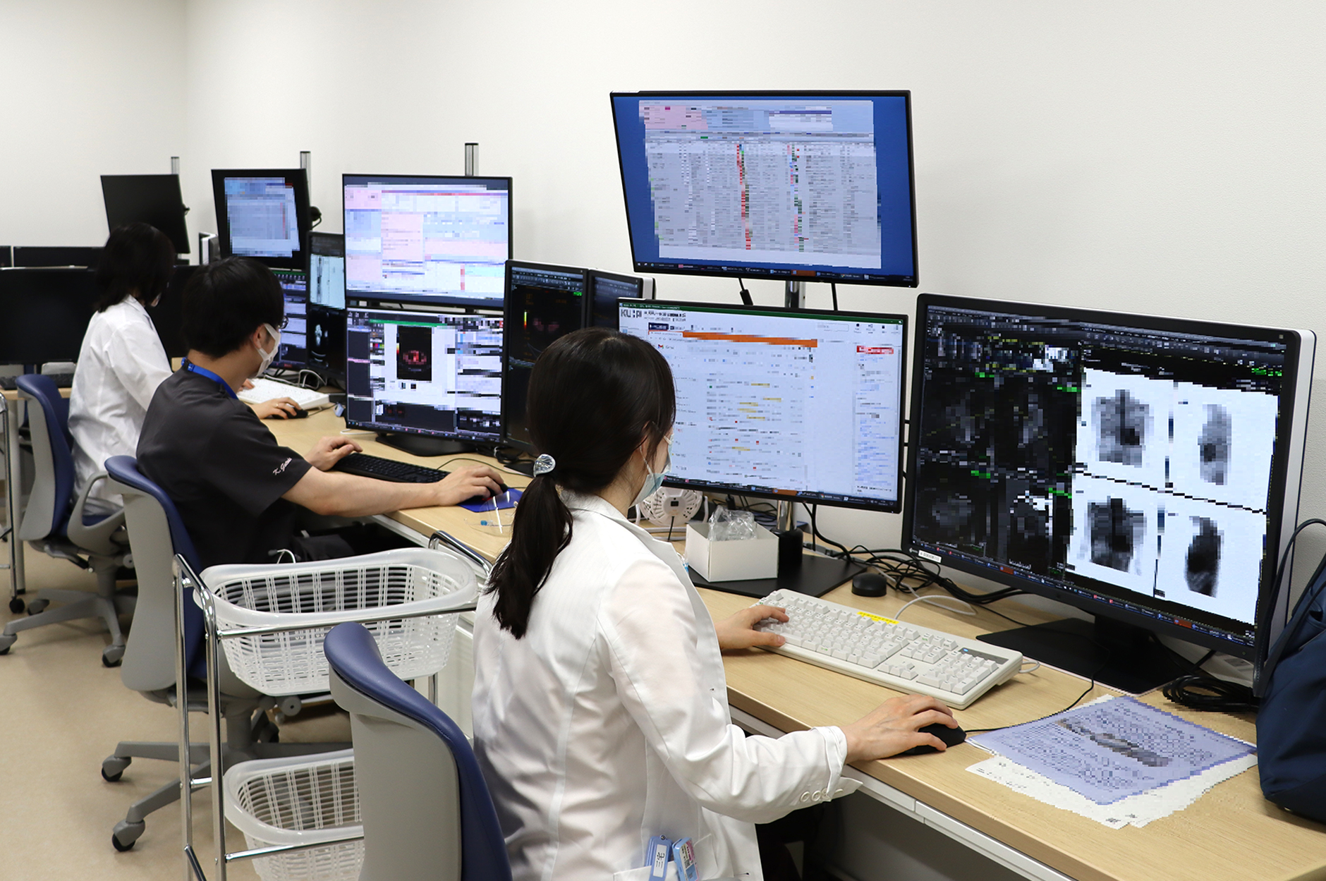
(Photo has been partially modified)
As a university with a mission to offer high-quality medical service, diagnostic imaging plays a crucial role for advanced treatments of malignancy and clinical trials of new drugs. It is important for radiologists to display pre-operative diagnostic images from multiple modalities simultaneously on monitors for comparison, to make final diagnoses, to determine the extent of lesion progression, and to respond to consultations from physicians with different specialties.
(Photo has been partially modified)
Why did you choose these monitors to add to your workflow?
Balanced Display of Diagnostic Image Data
When considering to purchase new monitors, we asked EIZO to show us several different models. Our choice was the 6-megapixel RadiForce RX660 color monitor for CT, MRI, RI, and angiography due to it having the best balance between space for the interpretation software menus and a 2x4 tiled view of the diagnostic images. For mammography we chose the 12-megapixel RadiForce RX1270 color monitor. These monitors are used in a three- or four-monitor configuration with standard FlexScan EV2456 monitors (1920×1200 resolution) to display patient information and diagnostic reports.
 (Photo has been partially modified)
High Quality Monitors are Underpinning High Quality Image Interpretations
When image interpretation was film-based, quality was determined by the performance of the imaging equipment, such as CT scanners. Today, image quality depends on size, brightness, and other characteristics of the monitors, so the quality of the monitor has a major impact on the quality of our reading.
It may sound like an exaggeration, but I believe that the quality of EIZO monitors ultimately determines the quality of our interpretations. I hope that EIZO will continue its work as a monitor manufacturer to enhance the standards for monitors throughout the industry.
What was your experience with the monitors?
Comparing Images to Spot Subtle Changes
In the past, we used to use two monitors in portrait orientation to do readings. Before starting to read images, there were all kinds of little adjustments we had to make – moving the monitors together to eliminate the gap between them, aligning their heights, and making sure they were both tilted the same. Now with a single wide monitor, we are free from these adjustments. I can start reading images as soon as I get to my desk. It contributes a lot in reducing stress.
The old monitors had quite thick bezels, which made image comparisons between the two tandem monitors awkward. Replacing them with a single RX660 monitor in landscape orientation has made comparisons much faster. Although the total screen area is the same as before, having a wide-screen format makes it much smoother to compare current and previous diagnostic images to spot subtle changes and new lesions. In a setting of clinical trials, comparison of detailed images in chronological order is mandatory. The RX660’s wide screen can display up to eight images acquired at different timepoints simultaneously.
Multi-Modality Image Comparisons
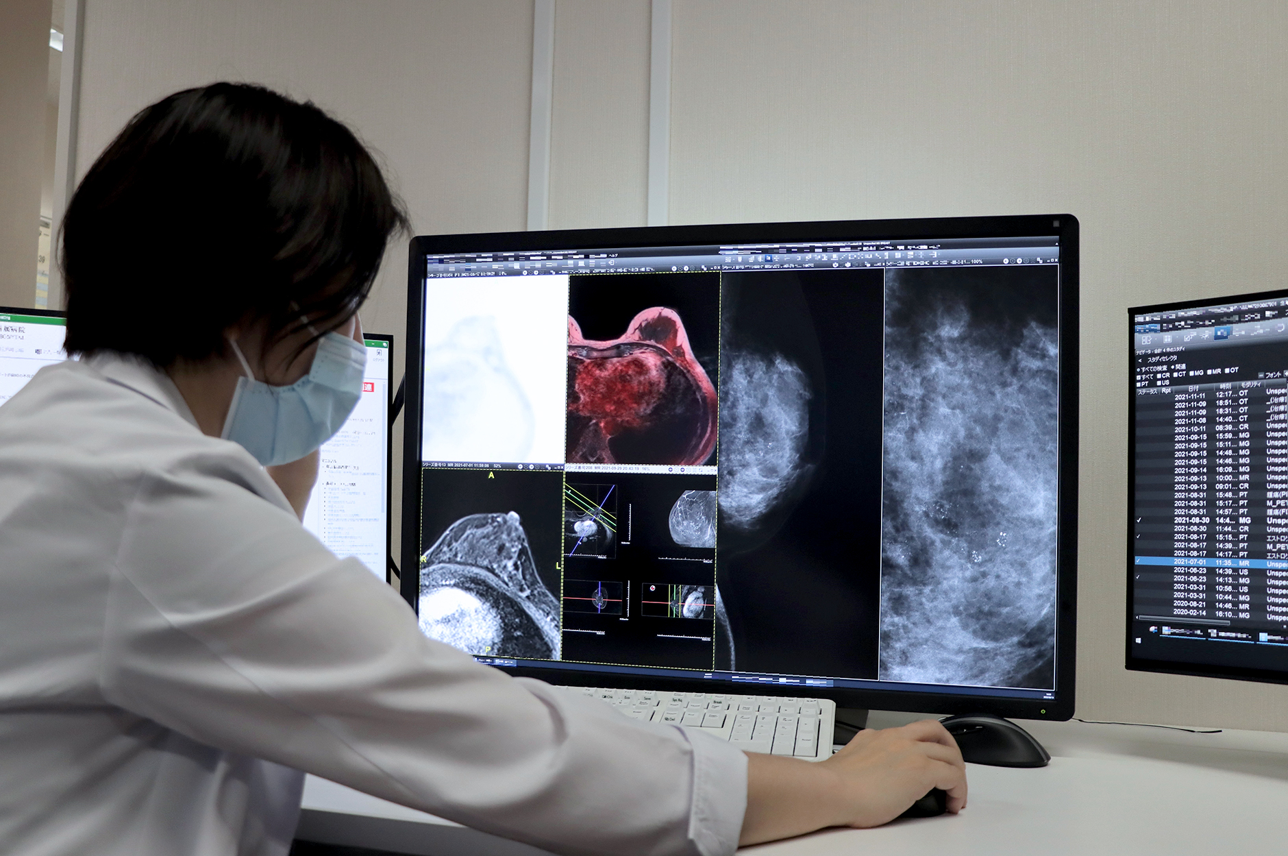
In breast imaging, mammography is the diagnostic imaging modality that demands both high contrast and high spatial resolution. Each pixel of the image represents just a few tens of microns, yet the appearance of a few or several microcalcification can differentiate malignant from benign breast lesions. It is critical for accurate diagnoses that a monitor can display at least 5-megapixel images pixel-perfect at 100% size.
On the other hand, for preoperative cases, it’s often best to do readings in conjunction with the patient’s breast MRI scans, PET scans, with the use of fusion imaging requiring color-overlay. These comparisons may often lead to new insights. The different diagnostic imaging modalities contribute in a complementary manner; having more eyes on a patient’s pathology means we can get more information and make more precise diagnoses, avoiding missing things.
Image reading used to be much less efficient in the past – we had to switch back and forth between high-resolution monochrome monitors for mammography images, and color monitors for MRI and PET scan images. The ability to display images from these different modalities on a single color monitor, the RX1270, has led to revolutionary improvements in efficiency.
Eyestrain Prevention
The matte surface of the screen significantly reduces reflections and glare, which is great for preventing eyestrain. I really appreciate that because we work with these monitors all day long. That’s also why we use EIZO monitors for our own research work.
What is your outlook for the future?
|
There is a growing need for monitors and software that can display various patient information at the same time. As the use of AI advances, we’ll need even more space to display data, so I expect that our current monitor setup may not be able to address that. It would be ideal to create a display environment that can show rich information in a limited space, not only within the Department of Diagnostic Imaging and Nuclear Medicine, but also for outpatient departments.
|
 |
![]()
 |
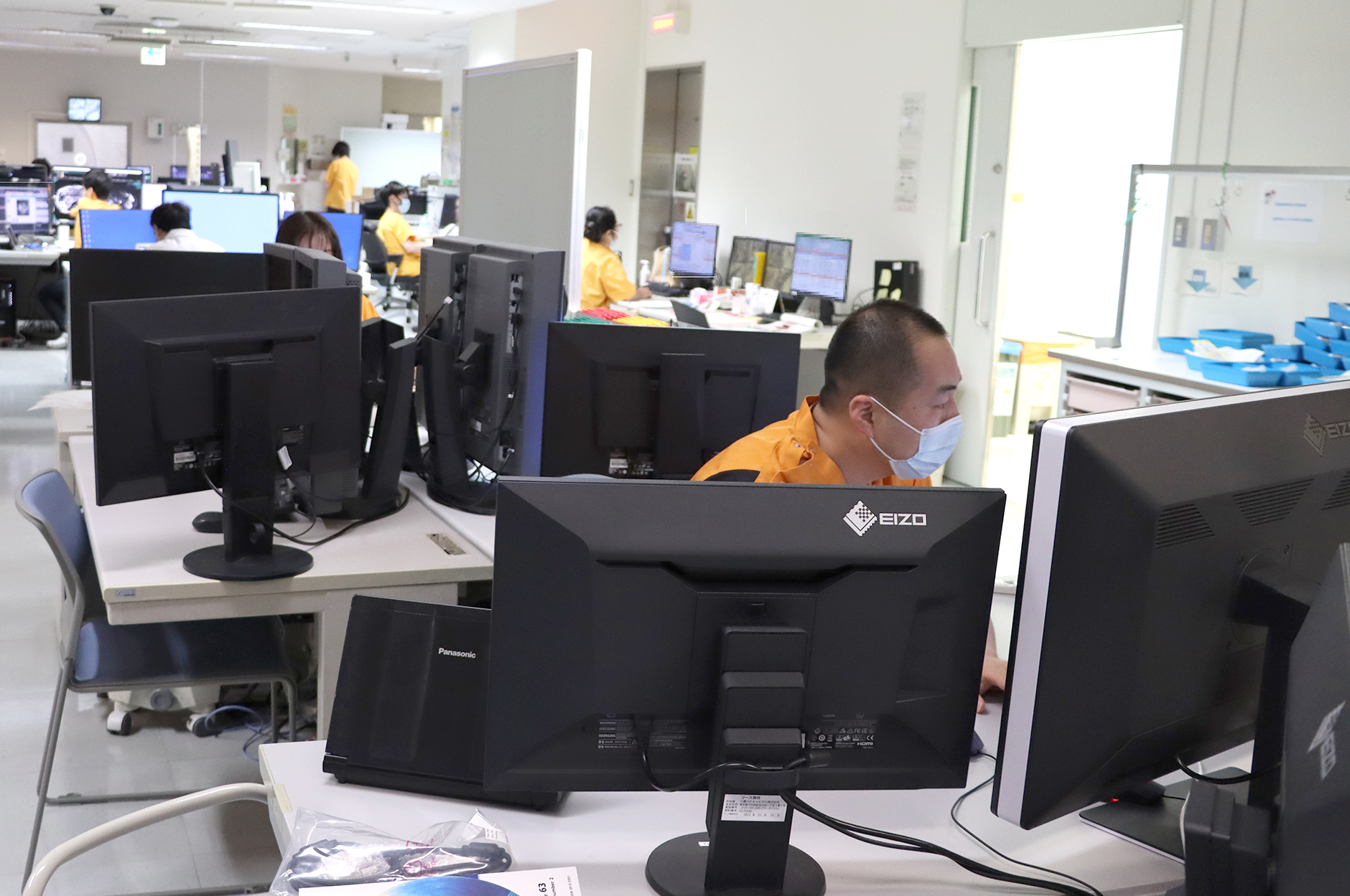
Kyoto University Hospital also uses EIZO monitors for displaying electronic health records. |
Kyoto University Hospital Website
https://www.kuhp.kyoto-u.ac.jp/english/
The photo shooting was conducted after taking infection control measures by hospital staff.